Center for Breast Care
Tomosynthesis and Contrast-Enhanced Mammography
Lexington Clinic offers the latest breast cancer detection technology with tomosynthesis and contrast-enhanced mammography.
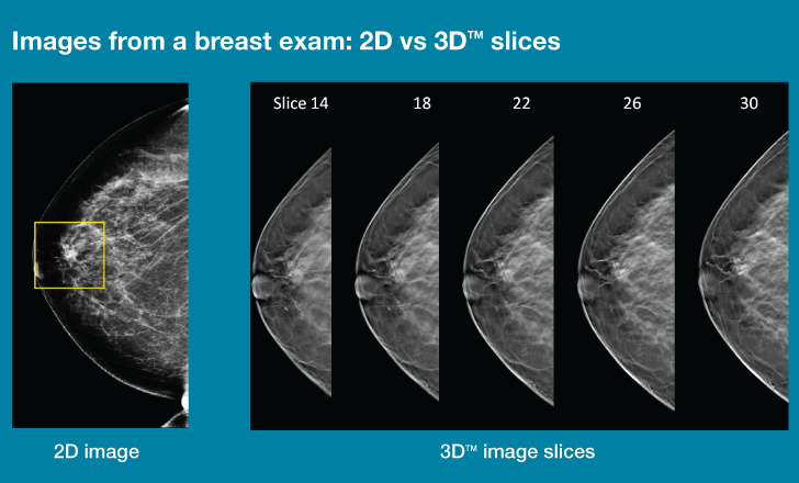
Tomosynthesis, also known as 3-D mammography, provides a better look at the breast than traditional testing. Tomosynthesis takes a series of images while rotating around the breast, giving doctors the ability to examine the area in "slices.”
Traditionally, providers have used 2-D images to look for breast cancer. This can cause problems when cancers are hidden by overlapping normal tissue. Tomosynthesis eliminates this issue by displaying the breast in slices and allowing providers to see any abnormalities.
Traditional 2-D screening mammography also has well-known limitations that require some women to come back for extra testing to rule out cancer. Tomosynthesis reduces the number of women who are called back for what ultimately turns out to be non-cancerous findings.
Lexington Clinic is now the first in Central Kentucky to offer contrast-enhanced mammography. This procedure uses a contrast injection similar to MRI to detect cancer.
Contrast-enhanced mammography is especially beneficial to women who have dense breast tissue and those with a family or personal history of breast cancer. Next to MRI, contrast-enhanced mammography is the most sensitive test available for detection of breast cancer because it lights-up tumors.
Contrast-enhanced mammography is test that has many of the advantages of breast MRI, including high sensitivity for cancer and a lower rate of false-positive results, compared with mammograms and breast ultrasound. It is also less expensive than standard breast MRI and takes less time.
To schedule your mammogram, please call +1 (859) 258-4444.

In a conventional 2D mammogram there appears to be an area of concern that the doctor may want to further investigate with another mammogram or a biopsy. Looking at the same breast tissue in slices from the 3D exam, the doctor can now see that the tissue is in fact normal breast tissue that was overlapping in the traditional mammogram creating the illusion of an abnormal area.

